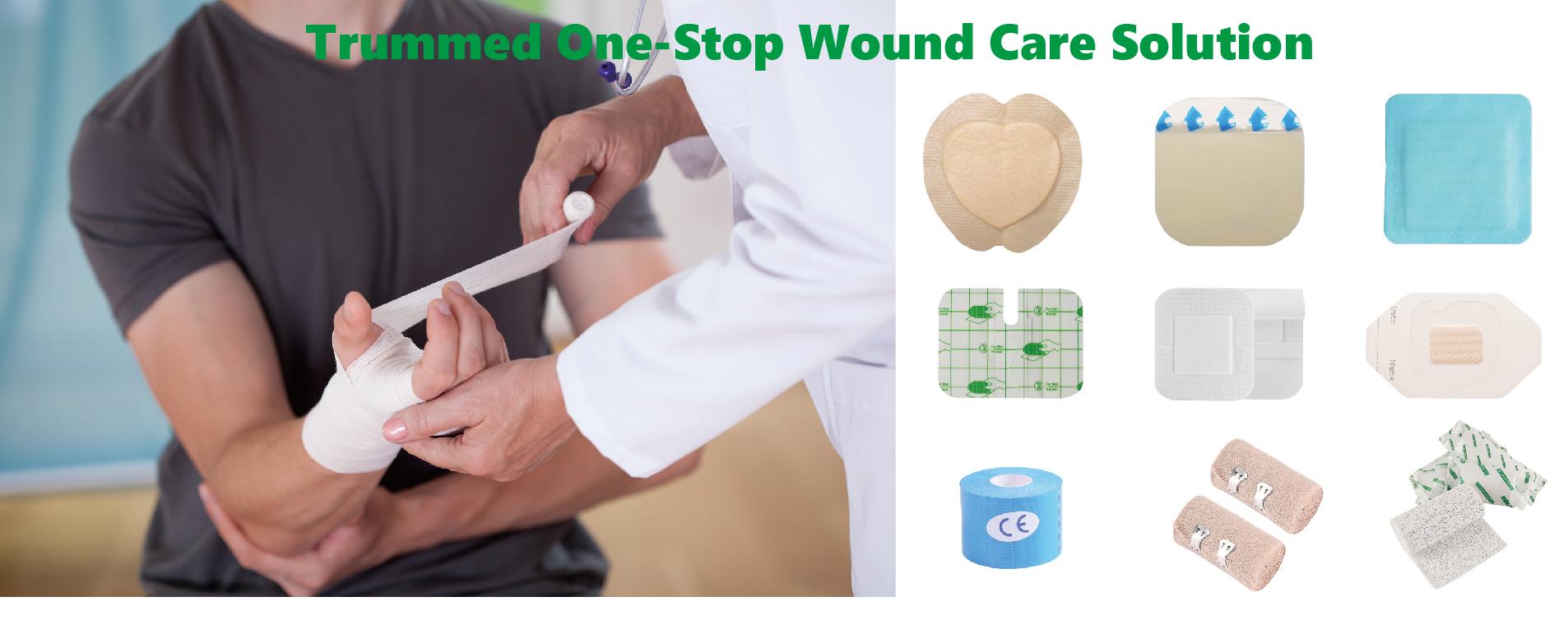
How to Choose the Suitable Wound Dressing
Choosing the right dressing to suit the conditions of a patient’s wound is vital for optimum healing and quality of life.
First Step: Asses your wound situation based on your following factors.
1) Wound measurement. Measure length, width, and depth in centimeters. Careful measurement of wound size is invaluable in evaluating the wound’s progress.
2) Appearance. Describe the color of the wound bed and assess the edges of the wound. These factors can help you determine the age of the wound, if healing has started, or if pressure or infection is present.
3) Exudate. Assess the color, amount, consistency, odor, and nature of wound drainage (exudate) before choosing a dressing.
4) Periwound tissue. Assess for signs of infection, such as erythema, edema, induration, warmth, crepitus, and damage from previous dressings.
Second Step: Asses your wound type.
1) Arterial wounds.
Atherosclerosis is the most common cause of arterial wounds. Other causes include trauma and thrombosis. Arterial wounds develop on the legs or feet distal to the narrowed or blocked artery.
2) Venous wounds
Venous wounds are almost the exact opposite of arterial wounds: Instead of getting too little blood, the legs have too much because the damaged vein valves can’t adequately return blood to the heart. Venous wounds often are large, with diffuse edges and yellow-white exudate.
3) Neuropathic wounds
Neuropathic wounds are most common over bony prominences and often occur on the foot below the ankle. The wounds are usually small and deep with thick callus formation at the wound edges (called hyperkeratosis).
4) Pressure ulcers
Pressure ulcers can be the most difficult and challenging wounds to care for. It’s important to remember that wounds covered with nonviable tissue can’t be staged.
Use this staging system that follows the recommendations of the National Pressure Ulcer Advisory Panel.
● Stage I—a defined area of persistent redness (in light-skinned patients) or persistent red, blue, or purple colors (in darker-skinned patients). The skin is intact, but compared with surrounding skin may be warmer or cooler, feel firm or boggy, and have altered sensation such as pain or itching.
● Stage II—a partial-thickness skin loss involving the epidermis or dermis and appearing as an abrasion, blister, or shallow crater.
● Stage III—a full-thickness skin loss including damage or necrosis of subcutaneous tissue. Damage may extend to, but not through, the fascia. Adjacent tissue may be undermined.
● Stage IV—a full-thickness loss with extensive skin damage, tissue necrosis, and possible damage to muscle, bone, tendons, or joint capsules. Sinus tracts and tunnels may be present.
5) Other wound types
surgical wounds, traumatic wounds and so on.
